KIMS-Arete Hospital spares no expense in enriching the department of neurosurgery to deliver humanistic and professional healthcare combined with the highest expertise and quality, ensuring the holistic healing of the patients.
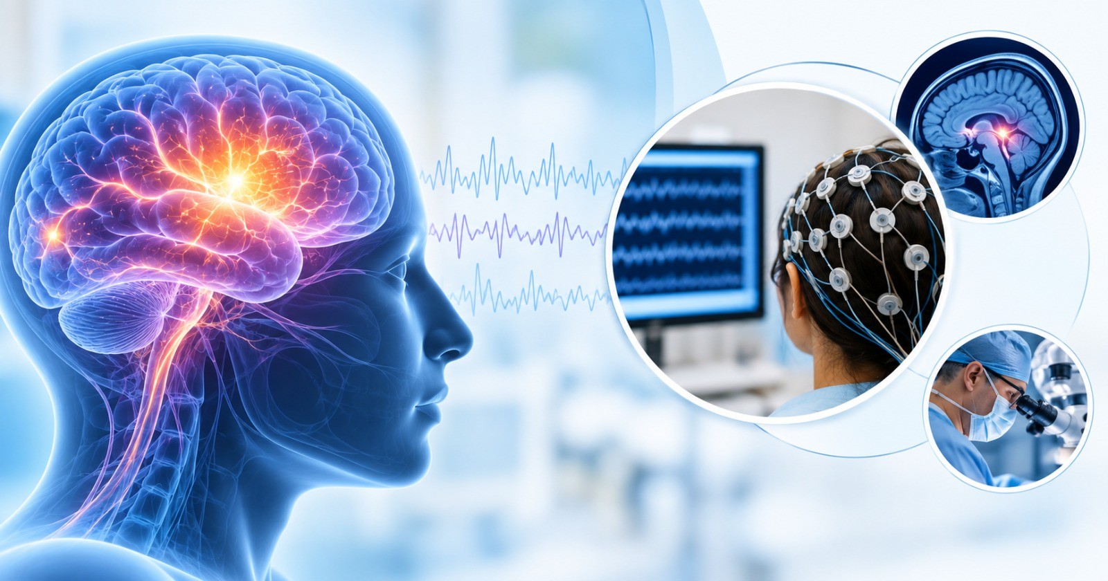
Being the best hospital for neurosurgery in Hyderabad, KIMS-Arete Hospital strives to be ahead of the curve, especially after understanding the fact that the coming generations shall bear witness to the disorders of the brain posing a great threat to humanity by capturing the highest mortality during their time. This is why KIMS-Arete Hospital heavily invests in the acquisition of novel neurodiagnostic equipment such as Neuronavigation Systems, Operating Microscope, Intraoperative Neuromonitoring System, Drill System, O-ARM etc which broadens the scope of neurosurgery, thus executing its role in reducing the neurological diseases.
One of the few medical institutions to hold a full-fledged functional neurosurgery department with the intent of reducing diseases of the brain and the spinal cord, KIMS-Arete Hospital cherry picks the best neurosurgeons who demonstrate not just the academic skill in their medical school but also integrate the skill of adapting and integrating the ever-changing medical technology through their years of practice.
Our distinguished team of neurosurgeons has extensive experience in the field of neuroanatomy and neurosurgery, whose proficiency is reflected in the perfect execution of deep, intricate neurosurgery procedures such as Awake Brain Surgeries, Microscopic Brain Surgeries, Skull Base Surgeries, Endoscopic Procedures, Cerebrovascular Procedures, among other procedures which are supported by meticulous Neurophysiological Monitoring.
Step in to obtain precise diagnoses for the conditions affecting the brain, spine, and spinal cord and comprehensive management arising from the conscientious, explicit, and judicious use of current best evidence in making decisions catered around patient-centric care.
Common Symptoms presented to Department of Neurosurgery:
- Headache & Vomiting: Headache is among the most common reasons; patients seek medical attention on a global basis. The duo of headache and vomiting could be one of the most common symptomatic presentations among many diseases, such as tumors, meningitis, subarachnoid hemorrhage, aneurysm, etc. In certain pathologies causing the increased intracranial pressure, headaches are presented, which can be treated with neurosurgery.[BM2.1]
- Sudden Loss of Consciousness: A sudden and temporary loss of consciousness is called syncope, commonly due to reduced blood circulation arising from neurologic or metabolic disorders such as intracranial bleed, stroke, etc., which may require neurosurgical intervention.[BM3.1]
- Progressive Loss of Vision: Visual loss describes a temporary or permanent reduction in visual acuity, which could arise from diverse factors. From a neurosurgical point of view, it could be due to various diseases that affect the optic nerve including brain tumours and pathologies causing increased intracranial pressure or any brain injuries.[BM4.1]
- Seizures: Sudden and uncoordinated electrical signal changes in the brain causing sudden stiffening and jerking of the arms and legs, or a loss of awareness or changed awareness level. Seizures which are refractory to medical management have a better chance of cure with surgery in selected cases.
- Weakness in Extremities: Weakness is often accompanied by fatigue, which could be due to various neuropathies. From a neurosurgical point of view, it could be pathology causing compression over brain/ spine tracts. Sudden onset of weakness could be due to stroke.
- Neck & Back Pain Radiating to Limbs: The neck and back pain could arise due to various neuropathies, (such as herniated disc compacting the nerve root, spondylosis, etc) which may require various neurosurgical interventions.[BM5.1] Rarely spinal cord tumors can present neck or back pain with or without radicular symptoms.
- Facial Pain and Spasm: One of the common causes of facial pain is trigeminal neuralgia, acute intermittent pain of the part of the face which is caused due to compression of trigeminal nerves at the brain stem. Microvascular decompression (MVD) is a common procedure to treat the ailment, comprising high success and low complication rates.[BM6.1]
- Gait Disturbances: A fundamental aspect of human movement, gait is the pattern of walking or running, and its disruptions can signal various underlying health issues. Gait disturbances are described as any deviation from normal walking or gait. These deviations occur in the intricate interplay of muscles, joints, and neurological pathways and present challenges that extend beyond mere physical inconvenience.[BM7.1] Gait disturbances could also be due to cranial or spinal pathologies or both, and to correct the same, a consultation with a qualified neurosurgeon might be necessary.
- Persistent Giddiness & Hearing Loss: Sudden hearing loss and vertigo have multifactorial causes, of which viral, autoimmune, and neurological are the most common.[BM8.1] Also, tumors originating from the 8th cranial nerve (such as Vestibular Schwannoma) can lead to gradual hearing loss due to nerve compression. Surgical interventions may be necessary to address these tumors and alleviate the symptoms.[BM9.1]
- New Onset of Speech Disturbances: Speech disturbances are common consequences of brain injury or neurodegenerative impairment. Sudden difficulties with speech may signal conditions such as cerebrovascular accidents, etc.[BM10.1] In some cases, progressive speech disturbances can also be related to the onset of tumors involving the speech area of the brain.
Subspecialities in Neurosurgery Department:
- Brain Tumours: Trained to navigate the complex and sensitive structures of the brain, the neurosurgeons at KIMS-Arete Hospital utilise the state-of-the-art equipment and infrastructure, along with their clinical skills, to address the unique challenges posed by each type of brain tumour.
- Spine Surgeries: Endoscopic or open spine surgeries can be catered to patients suffering from complex and challenging spinal conditions such as degenerative disc disease, herniated disc, lumbar spinal stenosis, scoliosis, spondylolisthesis, vertebral compression fractures, spinal tumours, traumatic spine surgeries, etc.
- Emergency Neurosurgery: KIMS-Arete Hospital caters to the neurosurgeries done at the need of the hour among patients suffering from intracranial bleeding, traumatic brain injury, among others, that necessitate timely diagnosis and rapid surgical intervention to prevent or reverse brain damage.
- Functional Neurosurgery: The restoration of neurological function, thus enhancing the quality of life among patients suffering from movement-related disorders, pain, spasms, and epilepsy with a patient-centric, and individualised approach.
- Paediatric Neurosurgery: The highly skilled team of neurosurgeons and anaesthesiologists is equipped with cutting-edge technology and state-of-the-art facility, including ultra-sophisticated operating theatres to ensure top-quality paediatric neurosurgery outcomes for both congenital and acquired conditions. With a compassionate approach and specialised expertise, we strive to optimise the well-being of children, fostering a supportive environment for families throughout the treatment journey.
- Neurovascular Interventions: The KIMS-Arete Hospital approach to surgical management involves a comprehensive multimodality team, including interventional neuroradiologists, conducting advanced endovascular neurosurgery with precision and enhanced efficiency. Aneurysm Clippings and Arteriovenous Malformation Excisions are a couple of the common microsurgeries that require extreme skill and precision. Procedures such as these involve the utilisation of Indocyanine green (ICG) filter in microscope which helps to know about the adequacy of clipping aneurisms without compromising the normal vasculature.
- Movement Disorders: The promise of comprehensive care extends to patients suffering from Parkinson's disease and other challenging movement disorders, utilising novel technologies such as deep brain stimulation (DBS) to provide personalised neurosurgery treatment plans tailored to each patient's unique needs.
Diagnostic Tests in Neurosurgery Department:
- Functional MRI (fMRI): This investigation maps the functional areas in the brain, helping the neurosurgeons to treat the pathology without disturbing the normal brain.[BM11.1]
- Diffusion tensor imaging (DTI): A relatively newer technology, diffusion tensor magnetic resonance imaging, is necessary for mapping the white matter of the brain, which works by measuring the diffusion of water molecules.[BM12.1] Through this test the neurosurgeons differentiate the tracts with pathology from those without, thus can perform surgery without disturbing them.
- Magnetic resonance spectroscopy (MRS): Magnetic resonance spectroscopy (MRS) is a noninvasive technique to measure the concentrations of various compounds in the brain. It can be used to differentiate gliomas from other diagnoses such as metastases, lymphoma, demyelination, oedema, necrosis, infection, etc.[BM13.1]
- Magnetic Resonance Angiography (MRA): MRA is an imaging diagnostic modality that uses a computer to synergise the magnetic field and radio waves to produce images of soft tissues, bones, and internal body structures. Brain MRA produces three-dimensional images of the blood vessels which are necessary for the detection of arterial stenosis (narrowing of the arteries), to rule out aneurysms (arteries bulging) and arteriovenous malformations.
- Computerised tomography (CT) scan: This imaging test uses X-rays to make detailed images of the body, depicting the details of the bones, muscles, fat, and organs from various angles. CT scans of the brain are necessary to diagnose conditions such as tumours or other lesions, injuries or acute trauma, bleeding in the brain, stroke, infections, etc.
- Digital Subtraction Angiography (DSA): This is a gold standard for angiography and it detects the blood flow by capturing images of blood supply in the brain. A catheter (a small, thin tube) is inserted into an artery in the leg and guided up to the arteries in the brain. A contrast dye is pumped into the catheter, and images of the arteries are obtained in the Cath lab.
Common Diseases and Conditions Requiring Neurosurgery:
Infectious Diseases
1) Meningeal tuberculosis (commonly called brain tuberculosis): This is an infectious disease that occurs when the bacterium - Mycobacterium tuberculosis infects the meninges (membranes surrounding the brain and spinal cord) or brain parenchyma. This is a life-threatening condition and can cause complications such as seizures, hearing loss, increased pressure in the brain, brain damage, stroke, etc.[BM16.1]
2) Meningitis: This is an infectious inflammation of the tissues which surround the brain and spinal cord that requires immediate medical care. While meningitis occurs due to various germs, bacterial meningitis is the most serious one, as it can be fatal within 24 hours. In various cases, sequalae of meningitis may be developed such as hydrocephalus. In such cases, surgical treatment is also initiated.[BM17.1]
3) Brain abscess: Caused due to various germs like bacteria or fungus. Surgical intervention may be necessary if the patients are non-responsive to medical management.
4) Spinal epidural abscess: This condition presents with back pain, fever, and sudden onset of weakness in limbs. Urgent neurosurgical intervention with pharmacotherapy is the gold standard in treating these patients and those who do not respond to antibiotics.[BM18.1]
Trauma
5) Head Injury and Trauma: Head injury that is caused by an outside force, which can range from a small, forceful bump, blow to an object entering the brain causing temporary or fatal problems.[BM19.1] External appearances do not necessarily provide the complete picture, neurosurgery consultation is always advisable especially if there is any loss of consciousness or bleeding from ear, nose or throat.
6) Vertebral Compression Fractures: The vertebrae, when subjected to pressure or trauma, develop small cracks and even breakages which weaken the spine. Surgical intervention is required when there is spinal instability or neurological deficit.
Spinal Disorders
7) Degenerative Disc Disease: Usually seen as we age, the degenerative disc disease is the deterioration or breakdown of one or more of the discs between the vertebrae of the spinal column, leading to pain, weakness, numbness, etc.
8) Herniated Disc: A common condition of the spine in which the disc present between the vertebrae is forced through a weakened part of the outer layer of the disc, thus compressing the nerves or spinal cord, causing pain and spinal cord dysfunction.[BM20.1]
9) Lumbar Canal Stenosis: One of the most important diseases demanding spinal surgery among the elderly, lumbar canal stenosis indicates the narrowing of the spinal canal in the lower back, causing significant disability.[BM21.1]
10) Cervical canal stenosis with myelopathy: Cervical stenosis denotes to the narrowed spine in the neck (cervical area). There are various causes. If a patient has congenital stenosis (narrowed spine in the neck since birth), they are much more prone to develop disc herniations or bone spur formation. The stenosis can then lead to spinal cord compression (myelopathy), which is usually painless.[BM22.1]
11) Scoliosis: An abnormal sideways spine curvature often diagnosed in adolescents, which can lead to cosmetic and functional concerns.
12) Spondylolisthesis: Condition of slipped vertebral body with respect to the adjacent vertebral body causing radicular or mechanical symptoms or pain.
13) [BM23.1] Craniocervical Junction Abnormalities: The craniocervical junction disorders are abnormalities of the bones that join the head and neck. They may be present at birth or arise due to injuries or disorders.[BM24.1] Sometimes surgical decompression, fixation, or both the procedures are necessary to treat the condition.
Functional Disorders
14) Epilepsy: Disturbed electrical activity in the brain depicted by seizures. It can turn life-threatening if the disease persists.[BM25.1]
15) Trigeminal Neuralgia: A condition characterized by pain originating from the trigeminal nerve, which starts near the top of the ear and splits in three, toward the eye, cheek, and jaw. The pain is described as stabbing-like, or electrical in sensation and could be so severe that the patients cannot eat or drink.[BM26.1]
16) Hemifacial Spasms: Hemifacial spasms are painless and involuntary spasms (twitching) of the face muscles, which usually happen due to the compression of root of the facial nerve by the cerebellar arteries.[BM27.1]
Tumours
17) Brain Tumours: Brain tumours are the diseases that are characterised by the development of abnormal cells in the brain. Brain tumours can form in any portion of the brain or skull, including the meninges (protective lining), the skull base (underside of the brain), the pituitary gland, brainstem, etc. There are about 120 different forms of brain tumours.[BM28.1]
18) Spinal Tumours: Abnormal growth of cells in the spinal cord. They can be primary (originating in the spine) or secondary (metastatic cancer cells arising from other cancers). Symptoms include pain, weakness, numbness, and walking difficulty.
Congenital Disorders
19) Hydrocephalus: This neurological disorder is characterised by an abnormal buildup of cerebrospinal fluid in the ventricles (cavities) within the brain, widening them and increasing pressure on the brain's tissues. This ailment could be congenital (present at birth) or could be from injury.[BM29.1]
20) Myelomeningocele (also called open spina bifida): This type of disease occurs as a birth defect in which the spine and spinal canal are not yet closed. Normally, it should be closed before birth. This ailment can be diagnosed during pregnancy. The necessary surgery (either within the womb or after birth) can be performed to repair the infant’s spine.[BM30.1]
21) Tethered spinal cord syndrome: This is a congenital neurologic disorder in which the spinal cord is attached to tissues, limiting its ascent within the spinal column leading to abnormal stretching of the neural tissue.[BM31.1]
22) Spina bifida: This birth defect occurs due to the incomplete formation of the spinal column, leaving a section of the spinal cord and spinal nerves exposed through an opening in the back.[BM32.1]
Vascular Disorders of the Brain
23) Vascular Brain Aneurysms: Aneurysm is an abnormal bulge that occurred due to a weakened area in the wall of a blood vessel causing abnormal widening or ballooning.
24) [BM33.1]Arteriovenous malformations: Abnormal fistulas that are formed between the arteries and veins. High arterial blood flow directly into venous structures can disrupt the venous walls and ultimately cause fatal haemorrhage in the brain.[BM34.1]
Emergencies in Neurosurgery
25) Stroke: Using cutting-edge surgical approaches such as thrombectomy and hemicraniectomy, our interdisciplinary team is committed to provide quick and thorough treatment for stroke. Our dedication to enhance the quality of life for concentrate not only on restoring the neurological function but also help in avoid long-term problems significantly.
26) Head Injury and Trauma: Increased pressure inside the skull and bleeding or clot development in the brain may result from head injury and trauma. Our very talented and experienced neurosurgeons at KIMS-Arete Hospital conduct emergency surgeries such as decompressive craniectomy, cranioplasty, craniotomy, and external ventricular drains to avoid secondary brain damage.