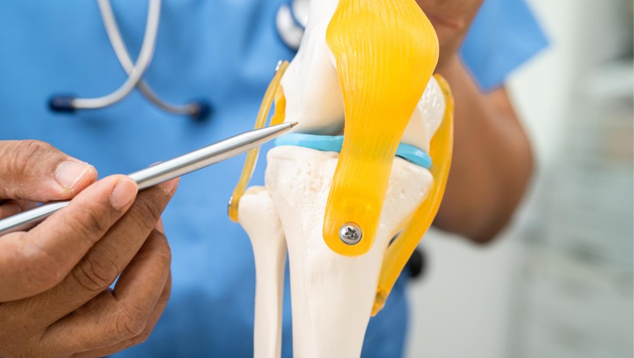
Cartilage Restoration for Chondral Lesions of the Knee
About Chondral Lesions of the Knee
Articular cartilage covers the bone ends inside the knee (femur, tibia, and patella) enabling frictionless movement and load distribution. When damaged, this tissue does not heal on its own. It has no blood supply. Nutrients reach it through joint fluid, which is insufficient for repair.
A chondral lesion, once formed, tends to enlarge over time. The surrounding cartilage destabilises, load distribution becomes abnormal and a contained focal defect progresses toward diffuse joint degeneration. Lesions are graded Grade I (surface softening) to Grade IV (full-thickness loss with exposed bone) using the ICRS classification. This grading, alongside lesion size and location, determines treatment.
Conditions included in chondral lesions of the knee are:
- Acute chondral and osteochondral injuries from sport or trauma
- Osteochondritis dissecans of the femoral condyle, trochlea, or patella
- Chondral lesions associated with ACL tears, meniscal injury, or patellofemoral instability
- Post-traumatic cartilage defects in younger patients not suitable for arthroplasty
- Symptomatic focal lesions identified on MRI.
Cartilage Restoration Procedures
Microfracture
An arthroscopic technique in which small perforations in the subchondral bone trigger a marrow-derived repair response. The tissue that forms is fibrocartilage - functional but inferior to native hyaline cartilage. Best suited for contained defects under 2 to 2.5 cm² in younger patients.
Autologous Chondrocyte Implantation (ACI) and Matrix-induced Autologous Chondrocyte Implantation (MACI)
A two-stage procedure using the patient's own cultured chondrocytes. A small cartilage biopsy is harvested, expanded in the laboratory over three to four weeks, then implanted into the defect on a collagen scaffold (MACI). Produces hyaline-like cartilage. Best suited for defects above 3 cm².
Osteochondral Autograft Transfer (OATS / Mosaicplasty)
Intact osteochondral plugs harvested from a low-load region of the patient's own knee are transplanted directly into the defect. Single-stage. Transfers genuine hyaline cartilage. Limited by donor-site availability and best for defects in the 1 to 4 cm² range.
Osteochondral Allograft Transplantation
For defects too large for autograft, osteochondral tissue from a screened donor is shaped to fit the defect and implanted in a single operation. The option of choice for large post-traumatic defects in patients not yet suitable for joint replacement.
Biologic Adjuncts
Platelet-rich Plasma and bone marrow aspirate concentrate are used selectively to improve the biological environment around a primary cartilage repair.
Rehabilitation
Non-operative management is considered first for lower-grade lesions. Surgery is recommended when the lesion profile or symptom burden makes it the appropriate path.
Rehabilitation is all about striking the right balance that is protecting the repair while restoring mobility and strength. The repaired cartilage needs progressive controlled loading to mature. It includes phased weight-bearing, range of motion exercises, muscle strengthening and guided physiotherapy. Recovery time includes:
- Microfracture: Return to sport four to six months.
- ACI / MACI: Return to full activity in twelve to eighteen months.
- OATS / Allograft: Variable; typically six to twelve months.
Why Choose KIMS Hospitals, Electronic City?
Not every orthopaedic department offers the full spectrum of cartilage restoration. KIMS Hospitals, Electronic City is a leading multispeciality hospital in South Bengaluru. Our Department of Orthopaedics and Sports Medicine manages the full range of joint and musculoskeletal conditions, with surgeons experienced in arthroscopic, joint preservation and cartilage restoration surgery. Our experts identify associated pathologies like ligament instability, malalignment, and meniscal deficiency and address them alongside the cartilage defect. Treating a lesion in a mechanically abnormal joint without correcting the underlying problem compromises the long term joint health. Rehabilitation is planned from the outset, not added after surgery. Patients receive a clear account of their imaging, their options, and realistic expectations.
Conclusion
Time is genuinely one of the most important factors when it comes to cartilage health and the longer a problem is left unaddressed, the fewer options remain on the table. The good news is that modern restoration techniques produce durable outcomes. Joints are preserved. Knee replacement is deferred.
At KIMS Hospitals, Electronic City, Bengaluru, you're supported through every stage of that journey from your very first consultation, through surgery, and all the way into rehabilitation. If you have a knee injury that has not resolved, unexplained recurrent swelling, or a chondral lesion on MRI, an early specialist review determines what options remain available. Waiting narrows them.
FAQs
What is a chondral lesion and why doesn't it heal on its own?
Articular cartilage has no blood supply. Without blood reaching the site of damage, the repair mechanisms that work for bone or muscle simply do not operate. A chondral defect does not close spontaneously and tends to worsen over time.
When should I see a specialist for knee cartilage damage?
Consult a joint specialist when you experience:
- Persistent pain in knee after a sports injury
- Knee swelling that returns without clear cause
- Mechanical symptoms like locking or catching
- An MRI mentioning a cartilage defect.
An earlier review preserves more surgical options.
What is the difference between microfracture and MACI?
Microfracture stimulates fibrocartilage repair from bone marrow. It is useful for smaller defects, but the tissue produced is mechanically inferior to native cartilage. MACI uses the patient's own cultured chondrocytes on a scaffold to regenerate hyaline-like cartilage. It is a two-stage procedure with a longer recovery but a superior biological outcome for larger lesions.
Can cartilage damage be treated without surgery?
Lower-grade lesions in patients with limited functional demands can be managed conservatively. This controls symptoms but it does not restore the tissue. Surgery is recommended when the lesion profile makes operative management the appropriate choice.
Does KIMS Hospitals Electronic City treat cartilage damage alongside other knee conditions?
Yes. In KIMS Hospitals Electronic City, our experts treat many knee conditions like ACL tears, meniscal injuries, and patellofemoral malalignment that frequently coexist with chondral lesions. Restoring cartilage without correcting associated mechanical problems reduces the durability of the repair.