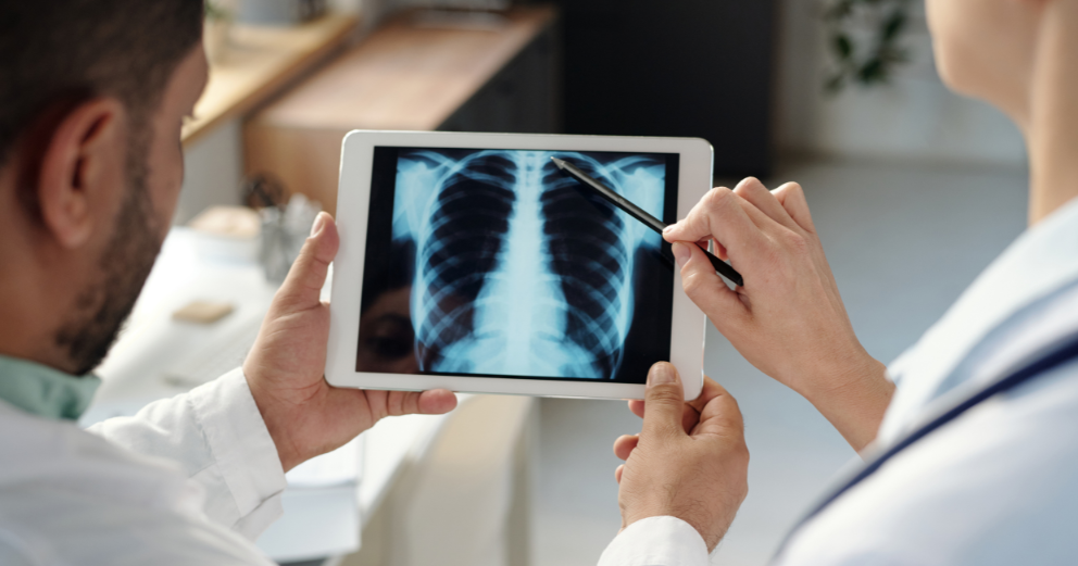
Radiology in Early Disease Detection
The value of catching a disease early is widely understood in principle. Treat cancer at Stage 1 and the outcomes are vastly better than at Stage 4. Identify heart disease before a heart attack rather than after. Find a clot before it causes a stroke. What is less widely appreciated is how central radiology is to making that early detection possible.
Imaging has moved well beyond its traditional role of confirming a suspected diagnosis. It now sits at the front line of disease prevention, screening programmes, and the identification of conditions that have not yet produced a single symptom.
Why early detection changes outcomes?
The relationship between early detection and better outcomes is most clearly demonstrated in cancer. By harnessing imaging modalities such as X-rays, CT, MRI, and PET, clinicians can visualise internal structures and detect abnormalities with unprecedented detail and precision. This non-invasive method allows for the early detection of diseases including cancers and cardiovascular conditions.
When cancer is found before it has spread beyond its site of origin, treatment is simpler, less aggressive, and significantly more likely to be curative. The same principle applies across conditions. Heart disease identified through imaging before a major cardiac event allows intervention that can prevent that event from occurring. Osteoporosis detected through a bone density scan before a fracture allows treatment that reduces fracture risk significantly.
Cancer screening through imaging
Several of the most effective cancer screening programmes in use today are built around specific imaging tests.
- Mammography for breast cancer
Mammography is the standard imaging tool for breast cancer screening. It uses low-dose X-ray to detect masses, microcalcifications, and architectural distortions in breast tissue, many of which would not be palpable for months or years. Regular mammography screening in women above 40 has consistently been shown to reduce breast cancer mortality by detecting cancers at earlier, more treatable stages.
- Low-dose CT for lung cancer
A radiomics-based reinforcement learning model to analyse serial low-dose CT scans has shown improvements in early diagnosis radiology of lung cancer at baseline screening. Low-dose CT of the chest is recommended for high-risk individuals, primarily long-term smokers above a certain age threshold, as an annual screening tool. It can identify small lung nodules that are undetectable on a chest X-ray and cannot be felt or suspected from symptoms at this size.
- Colonoscopy and CT colonography for colorectal cancer
Colonoscopy allows direct visualisation and removal of precancerous polyps before they develop into cancer, making it both a diagnostic and preventive tool. CT colonography, sometimes called a virtual colonoscopy, is an imaging alternative that uses CT to produce detailed images of the colon without the need for conventional endoscopy.
- MRI for prostate and cervical cancer
Multiparametric MRI of the prostate is increasingly used to guide decisions about biopsy and to characterise prostate lesions detected on PSA screening. In gynaecological oncology, MRI is used to evaluate the uterus and cervix when screening or symptoms raise concern.
Cardiovascular disease: imaging before the event
Radiology's role in early cardiovascular disease detection has grown considerably. Several imaging tools now allow the identification of cardiac risk before symptoms develop or a cardiac event occurs.
The coronary artery calcium scan is a low-dose CT that quantifies calcium deposits in the coronary arteries, providing a direct measure of atherosclerotic plaque burden. It is used in individuals with intermediate cardiovascular risk to refine risk stratification and guide decisions about preventive treatment. AI-driven coronary artery calcium scoring and aortic valve analysis from routine chest CT are among the advances in opportunistic cardiovascular risk assessment now being explored in clinical practice.
Coronary CT angiography provides detailed imaging of the coronary arteries and can identify significant blockages before they cause symptoms. It is increasingly being used as a first-line investigation for stable chest pain in preference to stress testing in many clinical settings.
Opportunistic screening: finding disease on imaging done for other reasons
One of the most significant developments in radiology is the concept of opportunistic screening, where findings relevant to other conditions are identified on scans performed for a different primary purpose.
A chest CT done to investigate a cough might reveal coronary artery calcium, vertebral fractures suggesting osteoporosis, or an incidental lung nodule. Opportunistic screening for incidental findings including pulmonary embolism, vertebral compression fractures, and osteoporosis detection from chest radiographs are all areas of active development in radiology. This approach extracts additional diagnostic value from investigations already being performed, without additional radiation exposure or cost.
The role of AI in early detection
Artificial intelligence is increasingly embedded in radiology workflows and is changing what can be detected and how quickly. AI technologies can assess imaging data at a scale and consistency beyond manual inspection, indicating small abnormalities that may be missed in high-volume reporting contexts.
In lung cancer screening AI helps to detect and characterise small nodules. In cardiac imaging, it assists in calcium scoring and plaque measurement. In breast imaging, AI-assisted mammography reading has shown gains in sensitivity, especially in the detection of malignancies in dense breast tissue. The clinical value lies not in replacing radiologists but in augmenting their ability to detect early disease consistently across large populations.
Takeaways
Radiology early disease detection has moved from a supportive role to a frontline one in preventive medicine. Imaging for cancer screening, including mammography, low-dose CT, colonoscopy and multiparametric MRI, detects malignancies before symptoms arise, when therapy is most successful. Early detection radiology in cardiovascular disease with coronary calcium scoring and CT angiography permits intervention before a cardiac event.
Opportunistic screening and AI-assisted image analysis are extending the reach of preventive imaging further than was previously possible. For patients with known risk factors for cancer or cardiovascular disease, understanding which imaging-based screening is appropriate for their age and risk profile is a conversation worth having with their doctor.