What Happens During an MRI?
An MRI is one of the most powerful diagnostic tools available in modern medicine, capable of producing highly detailed images of the brain, spine, joints, and soft tissues without using any radiation. And yet, for many patients, the prospect of having one produces more anxiety than almost any other routine medical procedure. The enclosed machine, the loud noise, the instruction to lie completely still for an extended period, none of these are particularly comfortable even when understood in advance.
Most of that anxiety comes from not knowing what to expect. A clear, honest account of what the MRI scan procedure actually involves tends to reduce it significantly.
How an MRI works?
A scanner for MRI employs radio waves and a strong magnetic field to produce detailed images of the interior of the body. The MRI scanner puts out signals that bounce off fat and water molecules in the body. A computer transforms these signals into clear images of organs, muscles, joints and other tissues. It is considered safe for most patients since it uses magnetism, not radiation, and may be repeated without the cumulative radiation issues of CT scanning.
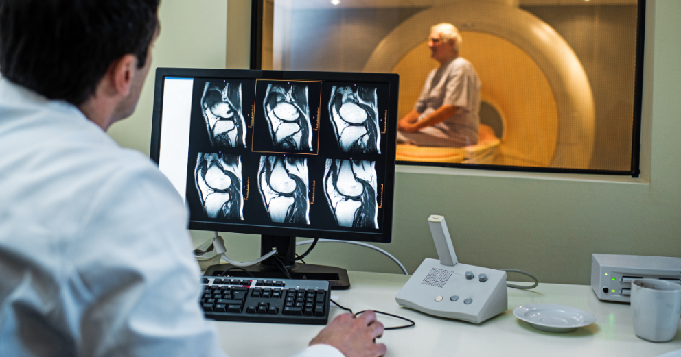
The scanner, itself, is a big cylindrical equipment with a central tunnel, commonly called the bore, in which the patient rests during the scan. The magnetic field generated is very powerful which is why metal objects and some implanted devices need thorough inspection before the scan can take place.
Before the scan: preparation
MRI Preparation begins before the patient even enters the scan room.
- All metal objects must be removed before entering the scanner area. This includes jewellery, watches, hair clips, belts, and any removable body piercings. Patients are usually asked to change into a hospital gown
- A safety screening questionnaire is completed to identify any implanted devices or metal in the body. Pacemakers, cochlear implants, certain joint replacements, and some other devices may be incompatible with the magnetic field and require discussion with the radiologist before the scan proceeds
- Patients are asked about allergies, particularly if contrast is being used, and about kidney function, since the contrast agent used in MRI is processed by the kidneys
- For abdominal or pelvic MRI, patients may be asked to fast for a few hours beforehand
- Anyone with claustrophobia should inform the team in advance, as several options are available to make the scan more manageable
If contrast is needed, a small cannula is placed in a vein in the arm before the scan begins. The contrast agent used in MRI is different from the iodine-based agent used in CT and is generally well tolerated.
During the scan
The patient lies on a narrow bed that moves into the MRI scanner, which is a long, narrow tube open at each end. Lying still on the table during the scan is essential, and scans typically take between 30 and 45 minutes to complete depending on the area being imaged.
Once positioned, the table slides into the scanner. The area of the body being examined is positioned within the central part of the machine where the magnetic field is strongest. Different areas of the body require different positions and coils, which are specialised devices placed around the relevant body part to improve image quality.
The most immediately noticeable feature of an MRI scan is the noise. The machine produces loud knocking noises during the scan. This is normal. Earplugs or headphones are provided to protect hearing. Different sequences are done to capture different tissue features and the sounds fluctuate in rhythm and strength during the scan.
The patient's job is simply to lie still while the machine takes images. Some patients describe the experience as relaxing. If uncomfortable at any point, it is easy to communicate with the technician, who is present throughout the entire scan. A hand-held buzzer or intercom system allows the patient to alert the radiographer immediately if they need to stop.
Managing claustrophobia
Patients often mention distress, anxiety, or claustrophobia related to MRI, resulting in missed appointments, disruptions to the scan, and lasting psychological effects for some. This is one of the most commonly raised concerns about MRI and is worth addressing directly.
Several approaches help patients manage claustrophobia during an MRI:
- Informing the team in advance allows them to walk the patient through the procedure step by step and ensure they feel as prepared as possible
- The scanner is open at both ends, meaning the patient can always see out, and the team is visible and reachable at all times
- Some centres offer open MRI scanners, which are less enclosed than standard machines, though they typically produce lower resolution images
- Music through headphones during the scan helps many patients feel less focused on the enclosed environment
- For patients with severe claustrophobia, a short-acting sedative can be arranged in advance through the referring doctor, allowing the scan to be completed without distress
After the scan
For most patients, there is no recovery period. Once the scan is complete, normal activities can be resumed immediately. If a contrast solution was given, the patient will be monitored for a few minutes to ensure there has been no rare allergic reaction to it.
A radiologist studies and reports the images. The findings are sent to the doctor who referred you. How long does this take? It depends on facility to facility and the urgency of the clinical issue. Emergency situations can be reported the same day or many days after routine scans.
Takeaways
During an MRI scan, a person lies within a giant magnetic scanner and detailed images of the area being scanned are made. This takes 30 to 60 minutes. Understanding what to expect out of an MRI scan, the noise, the tightness of the area, and the need to be still can minimise anxiety levels and improve the experience.
Before an MRI, you’ll need to remove all metal objects, fill out a safety screening form, and let the team know if you have any implanted devices or suffer from claustrophobia. MRI claustrophobia is a common problem and can be addressed by advance communication, proper setting, music or light sedation as necessary. It is a radiation-free treatment and is safe for most patients including those requiring frequent imaging.