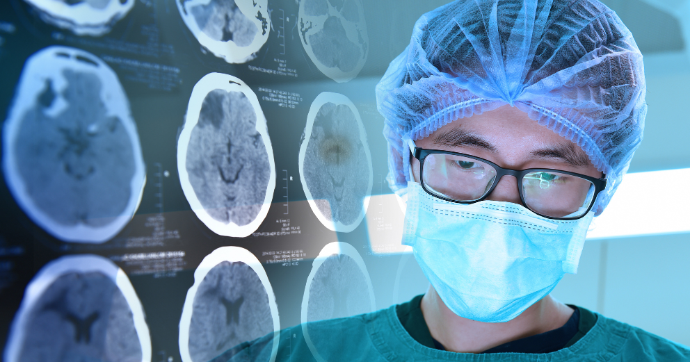
What Is Skull Base Surgery and When Is It Needed?
The skull base is the floor of the cranial cavity, the complex bony platform on which the brain rests. It is traversed by major blood vessels, cranial nerves, and critical anatomical structures in spaces so confined and so consequential that operating in this region has historically been among the most technically demanding work in all of surgery. Skull base surgery refers to the specialised surgical techniques developed to access, remove, or treat lesions in this region with precision and as little collateral disruption as possible.
What Makes the Skull Base Challenging?
The difficulty is anatomical. Tumours or other lesions at the skull base are adjacent to structures that cannot be easily sacrificed: the optic nerves, the carotid arteries, the brainstem, the cranial nerves controlling eye movement, facial sensation, swallowing, and hearing. Access through standard craniotomy approaches is limited by the geometry of the anatomy, and the depth of the operating field means that even minor errors in trajectory carry significant consequences.
Skull base surgery has been transformed over the past two decades by the development of endoscopic techniques that allow surgeons to approach tumours through the natural corridors of the nose and sinuses, reaching lesions at the centre of the skull base without any brain retraction at all.
Conditions That Require Skull Base Surgery
- Pituitary tumours are the most common reason for skull base surgery. The pituitary gland sits in a bony cavity called the sella turcica at the centre of the skull base. When a pituitary adenoma grows large enough to compress the optic chiasm or cause hormonal overproduction, surgical removal through an endoscopic transsphenoidal approach, through the nose and sphenoid sinus into the sella, is the standard treatment and avoids any external incision.
- Meningiomas arising from the meningeal coverings at the skull base, including the sphenoid wing, cavernous sinus, petroclival region, and olfactory groove, require removal through approaches tailored to the specific location and the structures involved.
- Acoustic neuromas, also called vestibular schwannomas, are benign tumours arising from the nerve that connects the inner ear to the brain. They cause progressive hearing loss, tinnitus, and balance disturbance, and when they grow large enough to cause brainstem compression, surgical removal through posterior fossa or middle fossa approaches is required.
- Craniopharyngiomas, chordomas, and chondrosarcomas are less common but similarly challenging tumours that arise in or around the skull base and require the specific surgical expertise that skull base surgery provides.
- Cerebrospinal fluid leaks and certain vascular malformations of the skull base are also managed through skull base surgical approaches.
What Patients Should Know
Skull base surgery is performed by neurosurgeons with subspecialty training in skull base surgery, often in collaboration with ENT surgeons for endoscopic approaches. Results are very dependent on the experience of the team, the technology available and the meticulous planning that precedes the procedure. Advanced MRI and CT imaging, intraoperative navigation, and neuromonitoring during surgery have all improved the safety and precision of these procedures considerably.
Conclusion
For patients facing a skull base diagnosis, the most important step is ensuring that the surgical team has specific experience with the condition and approach in question. The anatomy forgives very little, and expertise in this subspecialty is genuinely meaningful.